Posted on Leave a comment

Thymosin Alpha-1 Research Overview: 6 Proven Immune Peptide Mechanisms

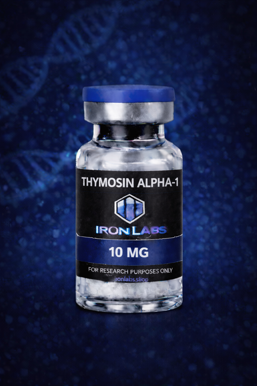
Thymosin Alpha-1 Research Overview - immune peptide vial

For research use only. All peptides referenced are research chemicals not approved by the FDA for human use. Not for human consumption.

This Thymosin Alpha-1 Research Overview examines Thymosin Alpha-1 (Tα1), a 28-amino-acid peptide fragment derived from the prothymosin alpha protein. Originally isolated from thymic tissue, Thymosin Alpha-1 has become one of the most widely referenced peptides in immune modulation research due to its role in T-cell maturation and broader immune signaling pathways.

Thymosin Alpha-1 Research Overview: Background & Discovery

Thymosin Alpha-1 was first characterized from thymic extracts as researchers investigated factors responsible for T-lymphocyte maturation. Since its isolation, it has been studied extensively as a reference peptide in immunology research, particularly regarding its influence on dendritic cell function and adaptive immune signaling cascades.

Research Areas & Mechanisms

This Thymosin Alpha-1 Research Overview highlights several mechanisms explored in laboratory literature:

  • T-cell maturation: Laboratory models have examined Thymosin Alpha-1’s role in promoting maturation and differentiation of T-lymphocytes within thymic tissue cultures.
  • Dendritic cell signaling: Cell culture research has explored its effect on dendritic cell activation and antigen presentation pathways.
  • Cytokine modulation: Research models have investigated Thymosin Alpha-1’s influence on cytokine profiles, including interferon and interleukin signaling.
  • Toll-like receptor research: Some laboratory studies have examined interactions between Thymosin Alpha-1 and TLR-mediated innate immune signaling.
  • Vaccine adjuvant research: Researchers have referenced Thymosin Alpha-1 in experimental models exploring immune response enhancement alongside vaccine antigens.

Across these research areas, Thymosin Alpha-1 is generally studied as a broad immune-modulating peptide rather than a pathogen-specific agent.

Thymosin Alpha-1 vs. LL-37 in Research Context

Thymosin Alpha-1 and LL-37 are both referenced in immune peptide research, but they represent different arms of the immune system. Thymosin Alpha-1 research centers on adaptive immunity, T-cell maturation, and dendritic cell signaling, while LL-37 research focuses on innate immune defense and direct antimicrobial activity. Researchers often reference both peptides together when comparing innate versus adaptive immune signaling models.

Laboratory Handling

Thymosin Alpha-1 is typically supplied as a lyophilized powder and should be stored at -20°C prior to reconstitution. Once reconstituted with bacteriostatic water, store refrigerated at 2-8°C and use within 14-21 days for research consistency. Avoid repeated freeze-thaw cycles and extended light exposure.

Source Thymosin Alpha-1 from Iron Labs

Iron Labs Thymosin Alpha-1 is supplied as lyophilized powder accompanied by third-party COA documentation, including HPLC purity and mass spectrometry identity confirmation, to support research reproducibility. Source Thymosin Alpha-1 for your research → Iron Labs Research Catalog

Frequently Referenced Research Questions

Is Thymosin Alpha-1 the same as Thymosin Beta-4? No. Thymosin Alpha-1 and Thymosin Beta-4 (the parent compound of TB-500) are structurally distinct peptides studied in different research contexts—immune modulation versus tissue repair, respectively.

Why is Thymosin Alpha-1 referenced in vaccine research? Researchers have studied its capacity to modulate immune responses alongside antigens in experimental adjuvant models, making it a recurring reference point in immunology literature.

This Thymosin Alpha-1 Research Overview will be updated as new laboratory literature becomes available to support ongoing immune peptide research.

Regulatory Notice

Thymosin Alpha-1 is not FDA-approved for any human or veterinary therapeutic application. Iron Labs sells Thymosin Alpha-1 exclusively as a research chemical for use by qualified researchers and laboratories. No health, therapeutic, or cosmetic claims are made or implied. Background on thymic peptide research is available via PubMed.

Researchers using this Thymosin Alpha-1 Research Overview as background often note that experimental dosing and timing protocols vary considerably across published laboratory models, which is why reviewing methodology sections closely remains an important step before designing comparable research.
Posted on Leave a comment

LL-37 Research Overview: 7 Proven Antimicrobial Peptide Mechanisms

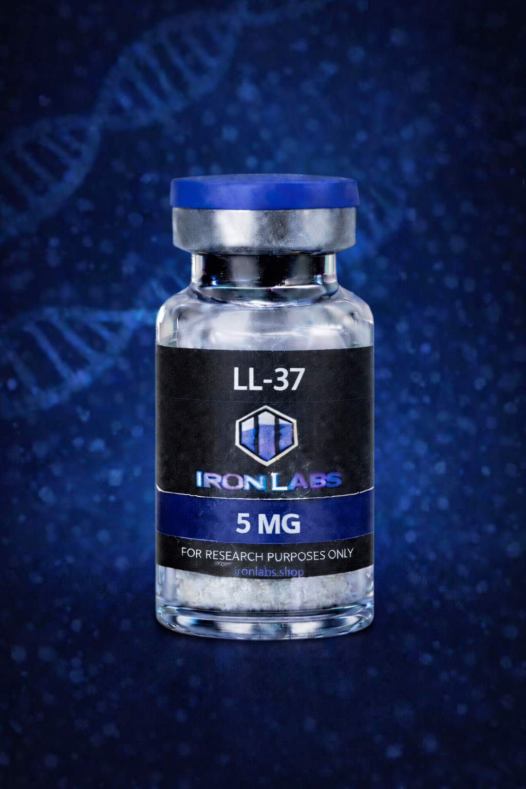
LL-37 Research Overview - cathelicidin research peptide vial

For research use only. All peptides referenced are research chemicals not approved by the FDA for human use. Not for human consumption.

This LL-37 Research Overview examines LL-37, the 37-residue human cathelicidin antimicrobial peptide derived from the CAMP gene product hCAP18. LL-37 is one of the most extensively studied peptides in innate immunity research due to its broad-spectrum antimicrobial activity and its role in modulating immune cell signaling.

LL-37 Research Overview: Background & Discovery

LL-37 was identified as the active peptide fragment cleaved from hCAP18, the sole human cathelicidin precursor protein. Researchers have studied its production by neutrophils, epithelial cells, and other immune cells, particularly in response to infection or tissue injury, making it a frequent reference point in innate immunity and antimicrobial peptide literature.

Research Areas & Mechanisms

This LL-37 Research Overview highlights several mechanisms explored in laboratory research:

  • Membrane disruption: In vitro studies have examined LL-37’s amphipathic helical structure and its capacity to disrupt bacterial membranes.
  • Biofilm research: Laboratory models have investigated LL-37’s effect on bacterial biofilm formation and established biofilm structures.
  • Immunomodulation: Cell culture research has explored LL-37’s role in chemotaxis, cytokine release, and modulation of both innate and adaptive immune responses.
  • Wound healing research: Animal and cell models have studied LL-37’s involvement in re-epithelialization and angiogenesis signaling.
  • Endotoxin neutralization: Some laboratory studies have examined LL-37’s interaction with bacterial lipopolysaccharide (LPS) and its downstream inflammatory signaling effects.

Across these research areas, LL-37 is generally studied as a multifunctional innate immune peptide rather than a single-mechanism antimicrobial agent.

LL-37 vs. Thymosin Alpha-1 in Research Context

LL-37 and Thymosin Alpha-1 both appear frequently in immune-related peptide research, but their study contexts differ. Thymosin Alpha-1 research centers on T-cell maturation, thymic signaling, and modulation of adaptive immunity. LL-37 research instead focuses on innate immune defense, direct antimicrobial activity, and epithelial barrier signaling, making the two peptides complementary reference points across innate versus adaptive immunology research models.

Laboratory Handling

LL-37 is typically supplied as a lyophilized powder and should be stored at -20°C prior to reconstitution. Once reconstituted with bacteriostatic water, store refrigerated at 2-8°C and use within 14-21 days for research consistency. LL-37 solutions should avoid repeated freeze-thaw cycles and extended exposure to light.

Source LL-37 from Iron Labs

Iron Labs LL-37 is supplied as lyophilized powder accompanied by third-party COA documentation, including HPLC purity and mass spectrometry identity confirmation, to support research reproducibility. Source LL-37 for your research → Iron Labs Research Catalog

Frequently Referenced Research Questions

Is LL-37 the same as hCAP18? No. hCAP18 is the precursor protein, and LL-37 is the active 37-amino-acid peptide fragment cleaved from it during immune cell activation.

Why is LL-37 studied alongside biofilm research? Researchers frequently reference LL-37 in biofilm literature because of its documented interactions with bacterial membrane structures in various in vitro models.

This LL-37 Research Overview will be updated as new laboratory literature becomes available to support ongoing antimicrobial peptide research.

Regulatory Notice

LL-37 is not FDA-approved for any human or veterinary therapeutic application. Iron Labs sells LL-37 exclusively as a research chemical for use by qualified researchers and laboratories. No health, therapeutic, or cosmetic claims are made or implied. Background on cathelicidin peptide research is available via PubMed.

Researchers evaluating this LL-37 Research Overview alongside their own literature review often note that concentration-dependent effects are a recurring theme: lower concentrations tend to favor immunomodulatory signaling in laboratory models, while higher concentrations are more frequently associated with direct membrane-disruptive antimicrobial activity in vitro. This dose-context distinction is a common consideration in experimental design for researchers working with cathelicidin-derived peptides.

Posted on Leave a comment

KPV Research Overview: Anti-Inflammatory Peptide Science & Laboratory Applications

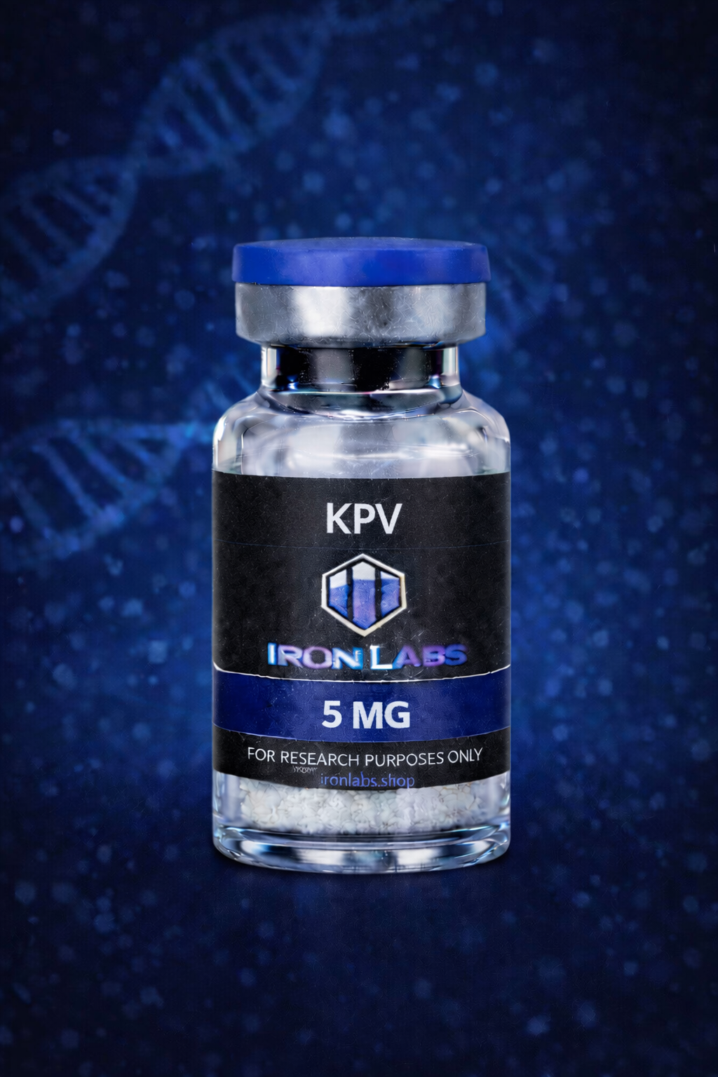
KPV Research Overview - 5mg lyophilized research peptide vial

For research use only. All peptides referenced are research chemicals not approved by the FDA for human use. Not for human consumption.

This KPV Research Overview examines KPV (lysine-proline-valine), the C-terminal tripeptide fragment of alpha-melanocyte-stimulating hormone (α-MSH), first identified for retaining much of the parent hormone’s anti-inflammatory activity without its pigmentation-related effects. Because of its small size and stability, KPV has become a frequent subject of in vitro and animal-model research into inflammatory signaling, skin barrier biology, and gut mucosal research models.

KPV Research Overview: Background & Discovery

KPV was identified as researchers investigated which fragments of α-MSH retained biological activity relevant to inflammation without triggering melanocortin receptor-driven pigmentation effects. This tripeptide fragment demonstrated that a relatively small sequence could still meaningfully engage inflammatory signaling pathways in laboratory models, making it an efficient candidate for researchers studying structure-activity relationships in melanocortin-derived peptides.

Research Areas & Mechanisms

This KPV Research Overview highlights several biological pathways that laboratory studies have explored:

  • NF-κB pathway modulation: Cell culture studies have examined KPV’s capacity to inhibit NF-κB nuclear translocation, a central transcription factor in inflammatory cytokine production.
  • Cytokine signaling: Research models have looked at KPV’s effect on IL-1β, IL-6, and TNF-α expression in stimulated cell lines.
  • Mast cell and mucosal research: Laboratory studies have investigated KPV in intestinal mucosal models, examining barrier integrity and local inflammatory markers.
  • Skin barrier studies: In vitro dermal models have explored KPV’s interaction with keratinocyte inflammatory signaling.
  • Oxidative stress research: Some laboratory models have examined KPV alongside markers of oxidative stress in inflamed tissue cultures, though this remains an early area of study.

Across these research areas, KPV is generally studied as a signaling modulator rather than a structural repair agent, distinguishing it from peptides more commonly associated with direct tissue remodeling.

KPV vs. BPC-157 in Research Context

KPV and BPC-157 are both frequently referenced in inflammation and tissue-repair literature, but their proposed mechanisms diverge substantially. BPC-157 research centers on angiogenesis, nitric oxide signaling, and FAK-paxillin pathway activity relevant to musculoskeletal and gastrointestinal models. KPV research instead concentrates on direct suppression of inflammatory transcription factors and cytokine cascades, making it a common reference point in immunology and dermatology-adjacent laboratory work rather than structural tissue repair studies.

Laboratory Handling

KPV is typically supplied as a lyophilized powder and should be stored at -20°C prior to reconstitution. Once reconstituted with bacteriostatic water, store refrigerated at 2-8°C and use within 14-21 days for research consistency. Avoid repeated freeze-thaw cycles, which can degrade peptide integrity, and protect from prolonged light exposure.

Source KPV from Iron Labs

Iron Labs KPV is supplied as lyophilized powder accompanied by third-party COA documentation, including HPLC purity and mass spectrometry identity confirmation, to support research reproducibility. Source KPV for your research → Iron Labs Research Catalog

Regulatory Notice

KPV is not FDA-approved for any human or veterinary therapeutic application. Iron Labs sells KPV exclusively as a research chemical for use by qualified researchers and laboratories. No health, therapeutic, or cosmetic claims are made or implied. Background on melanocortin-derived peptide research is available via PubMed. This KPV Research Overview is intended solely for laboratory and research reference purposes.

Frequently Referenced Research Questions

Is KPV the same as alpha-MSH? No. KPV is a small tripeptide fragment derived from the C-terminal end of alpha-MSH, retaining select anti-inflammatory signaling properties without the full hormone’s melanocortin receptor-driven pigmentation effects.

How is KPV typically studied alongside other peptides? Researchers frequently reference KPV alongside other immune- and inflammation-focused peptides to compare transcription factor targets, cytokine profiles, and downstream signaling outcomes across different experimental models.

This KPV Research Overview will be updated as new laboratory literature and research summaries become available, supporting researchers who require current background before designing their own studies.